


A family of five is exposed to the flu virus, but only two of them get sick. A lifelong chain-smoker lives 100 years, while a professional athlete in their prime dies of an unexpected heart attack. A drug fails a clinical trial due to harmful side effects in a subset of patients despite showing good results in others.
Could any of these scenarios have been predicted? The simple answer is not yet – but perhaps soon with the help of biomarkers.
A biomarker is anything measurable in the body that gives clinicians insight into someone’s current or future health. Biomarkers come in many forms and play a critical role in modern medicine by shedding light on the inner workings of the human body.
Many biomarkers are measured during routine procedures at the doctor’s office, including through blood tests and vital signs.
Physiological biomarkers such as heart rate and blood oxygen levels indicate how well your body is functioning. Cellular biomarkers such as white blood cell count provide useful information on the activity of your immune system. Genetic biomarkers are parts of your DNA that provide clues about your inherent risk of developing a particular disease or how likely you are to respond to a treatment.
There are still many more biomarkers to be discovered, and researchers are devising unique ways to identify a person’s risk of disease.
Discovering new biomarkers
The traditional way researchers identify biomarkers is by asking which biological features differ between two or more groups. This might look like comparing patients with a specific disease with healthy volunteers, people who respond to a drug with people who don’t, or the same group of people at two different points in time.
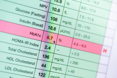
Many biomarker discoveries started with observations made in laboratories. For example, doctors use hemoglobin A1c levels in the blood to test for diabetes. The origins of this test date back to the 1960s, when scientists found abnormally high levels of a protein in the blood of diabetics. Repeated testing in the clinic over the next few decades confirmed the relationship between A1c levels and diabetes, culminating in the Food and Drug Administration approving the first A1c-based diabetes diagnostic test in 2013.
Other biomarkers that also started with observations made through basic research in the lab include screening tests for colorectal cancer, drug metabolism and inherited metabolic diseases in newborns.
Genetic biomarkers are typically found by scanning the genomes of thousands to even millions of people for genetic variants linked to a trait or disease. This approach is called a genomewide association study. This method has resulted in many important biomarkers that provide guidance on how to manage a patient’s risk of heart disease, age-related vision loss and liver disease.
One limitation of using the genomewide association study, though, is that it does not always provide information on which tissue or cell types may be relevant to the genetic variant. This lack of cellular information could be solved with cell cultures that allow researchers to study specific cell types under controlled conditions outside the body.
Human populations in a dish
In my work as a neuroscientist and geneticist, I take a different approach to finding biomarkers in the lab: I conduct genomewide association studies on human brain cells in a dish to understand how cells change when they encounter harmful exposures such as toxins and viruses.
Cells are the building blocks of all human tissues and organ systems. How sensitive they are to different environmental factors can greatly affect a person’s health and disease risk.
Thanks to revolutions in stem cell technologies, scientists are now able to model the effects of the environment on many different cell types – including brain cells that are nearly impossible to study in living people without causing irreversible damage to the brain. Instead, my colleagues and I obtain stem cells created from the blood or skin of human donors and convert them to brain cells for our experiments.
Most studies focus on how cells from only a handful of people respond to environmental stressors, mainly because it is quite difficult to conduct experiments at the population scale in a petri dish. To know whose cells are in a dish, researchers traditionally needed to handle and maintain cells from different people separately. This requirement adds considerable time, effort and cost for every new cell line included in the experimental design.
Because these issues limit the scale of experiments, studies aren’t able to capture the natural diversity of people and therefore cannot answer questions about why an exposure might make one person sick but have no effect on another.
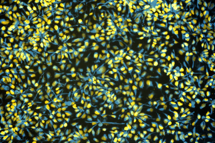
My laboratory tackles this problem by pooling stem cell-derived brain cells from dozens to hundreds of people into the same culture dish – what my team calls a cell village. We use the genomes of these cells as natural barcodes to determine who donated each one. We then add different environmental perturbations to the village – such as pathogens, chemical pollutants and radiation – and measure how each person’s cells reacts.


Each cell might respond by turning on or off a set of genes, showing an increased or decreased propensity to die, or do nothing at all. Identifying which genes are linked to these particular responses can help explain why a person’s cells respond the way they do.
These genes are essentially biomarkers for the vulnerability of a specific cell type to an exposure.
An atlas of human vulnerability
While cell villages are relatively new, they have already helped researchers better understand what makes people different from each other.
For example, my team and I used cell villages to identify a biomarker that indicates a person may be particularly vulnerable to Zika virus infection. Zika is a pathogen spread by mosquitoes that can cause neurological disorders and microcephaly – small head size – in developing fetuses. Using stem cell-derived brain cells from 40 people, we found that not everyone was equally susceptible to infection. For some, fewer than 1% of their cells were infected by Zika virus; for others, it was greater than 99%.

Through additional cell village experiments, we discovered a genetic variant that regulates a powerful antiviral gene called IFITM3. In this case, the cells of a person with version A of this gene were more vulnerable to infection than the cells of a person with version B. Researchers have found links between this same genetic variant and COVID-19 and influenza A infection severity, suggesting it could be a genetic biomarker that can predict how certain people may respond to certain types of pathogens.
My team has also conducted similar work to identify genetic biomarkers for a person’s sensitivity to lead, a pervasive neurodevelopmental toxin. We are also using cell villages to find biomarkers for a person’s vulnerability to PFAS, also known as forever chemicals; measles virus infection; and stress hormones.
Our goal is to build what we call an atlas of human vulnerability: a catalog of genetic biomarkers to predict how each person might respond at the cellular level to a wide range of harmful exposures.
The future of biomarkers
The cell village is only one of many emerging technologies that could usher in a new era of biomarker detection.
As DNA sequencing costs continue to drop, whole genome sequencing could become more routine in the clinic and potentially improve the ability to find links between genes and health outcomes that healthcare teams can intervene in.
Researchers are developing AI tools to survey medical imaging such as CT and MRI scans with the intention of finding new biomarkers for cancer progression that would have likely gone unnoticed by the human eye.
Cellphones and smartwatches may also be useful in efforts to discover new health-related biomarkers. For example, some studies have used these and other wearable devices to find behavioral biomarkers that predict major depressive episodes.
The goal now is to test how well these biomarkers work in the real world and make them widely available so that doctors everywhere can have as much information as possible about their patients when making life-changing decisions.
This article is republished from The Conversation, a nonprofit, independent news organization bringing you facts and trustworthy analysis to help you make sense of our complex world. It was written by: Michael F. Wells, University of California, Los Angeles
Read more:
- Tenacious curiosity in the lab can lead to a Nobel Prize – mRNA research exemplifies the unpredictable value of basic scientific research
- I teach doctors how to interpret test results – and the labels ‘normal’ and ‘abnormal’ often hide what matters most
- PFAS leave fingerprints in your blood – researchers are figuring out how forever chemicals transform in your body to read these clues
Michael F. Wells receives funding from the National Institutes of Health, California Institute for Regenerative Medicine, Burroughs Wellcome Fund, Rose Hills Foundation, and Simons Foundation.


(0) comments
Welcome to the discussion.
Log In
Keep it Clean. Please avoid obscene, vulgar, lewd, racist or sexually-oriented language.
PLEASE TURN OFF YOUR CAPS LOCK.
Don't Threaten. Threats of harming another person will not be tolerated.
Be Truthful. Don't knowingly lie about anyone or anything.
Be Nice. No racism, sexism or any sort of -ism that is degrading to another person.
Be Proactive. Use the 'Report' link on each comment to let us know of abusive posts.
Share with Us. We'd love to hear eyewitness accounts, the history behind an article.